Recently, the Brain Imaging Laboratory led by Prof. Tian Qiyuan from the School of Biomedical Engineering (BME), Tsinghua University, published an online research paper titled "Effects of diffusion MRI spatial resolution on human brain short-range association fiber reconstruction and structural connectivity estimation" in Imaging Neuroscience—a top-tier journal in the field of neuroimaging. The study systematically evaluated the impact of diffusion magnetic resonance imaging (dMRI) strategies on the reconstruction of short-range association fibers (SAFs) in the human brain and constructed the first atlas of SAF proportion across brain regions.
Zheng Jialan, a 2025 Ph.D. candidate from BME, is the first author of the paper. The corresponding authors are Associate Professor Tian Qiyuan from BME, Tsinghua University, and Dr. Li Ziyu, a postdoctoral researcher at the University of Oxford (former undergraduate graduate of the Department of Biomedical Engineering, Tsinghua University, Class of 2021). Collaborators include scholars from multiple institutions such as Tsinghua University's School of Biomedical Engineering, University of Oxford, Harvard University, Stanford University, and University of California, San Francisco (UCSF).
Click here to view more information about the paper
Key Background and Innovations
Short-range Association Fibers (SAFs), as critical white matter pathways connecting adjacent gyri, are essential for efficient information transmission between brain regions. They are also among the earliest structures affected in neurodegenerative diseases such as Alzheimer's disease. However, primarily distributed within the superficial white matter layer (only ~1.5 mm thick), SAFs are highly susceptible to partial volume effects (PVE) in conventional-resolution dMRI, leading to signal masking by surrounding tissues and hindering accurate reconstruction and quantitative analysis.
The study innovatively utilized the gSlider sequence to acquire multi-resolution dMRI datasets (isotropic voxel resolutions of 0.96, 1.5, and 2 mm) and retrospectively downsampled data from public dMRI databases, systematically comparing the effects of various imaging strategies on SAF reconstruction.
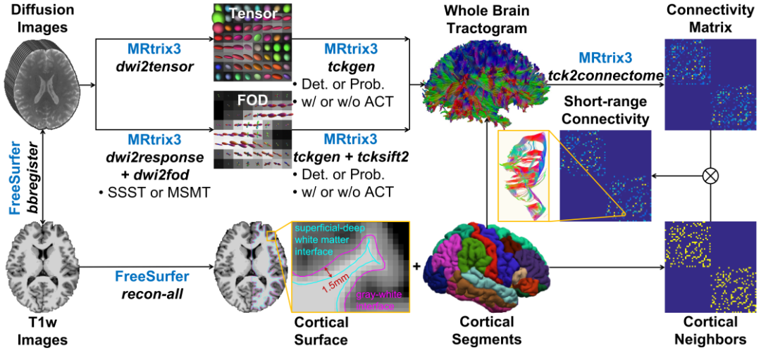
Figure: Research workflow
Major Findings
Lower spatial resolution significantly underestimates the proportion of SAFs in brain structural networks, with the temporal lobe being the most affected region.
Multi-fiber models based on Constrained Spherical Deconvolution (CSD), combined with anatomical constraints (ACT) and streamline filtering (SIFT), exhibit stronger robustness to resolution degradation.
Upsampling low-resolution data can partially improve reconstruction performance, particularly for Diffusion Tensor Imaging (DTI) methods.
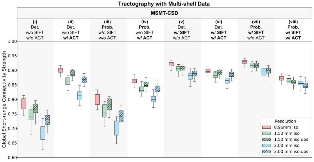
Figure: Proportion of SAFs in brain connectivity across different imaging methods
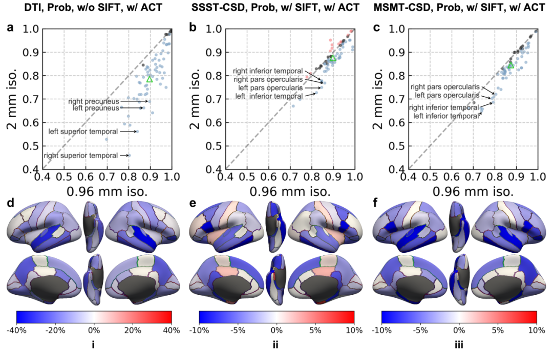
Figure: Sensitivity of SAF reconstruction proportion to resolution in various brain regions
Based on 0.96 mm high-resolution data and an optimized tracking pipeline, the study constructed the first atlas of SAF proportions across human brain regions, further confirming the dominant role of SAFs in brain connectivity and providing a new benchmark tool for understanding brain connection architecture.
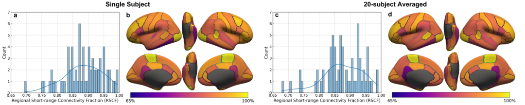
Figure: Atlas of SAF proportions in different human brain regions
Significance
This research provides systematic quantitative evidence for the selection of dMRI data acquisition and analysis methods, holding significant value for advancing the application of SAFs and connectomics in basic research and clinical disease diagnosis/treatment.
Introduction to Tian Qiyuan's Research Group & Brain Imaging Laboratory
Founded in 2023, the Brain Imaging Laboratory led by Prof. Tian Qiyuan in the School of Biomedical Engineering, Tsinghua University, is dedicated to driving the development and breakthroughs of new brain imaging tools, novel brain science discoveries, and innovative diagnosis/treatment strategies for brain diseases. The laboratory focuses on the research and development of artificial intelligence-enabled biomedical imaging and image analysis technologies, with core interests in cutting-edge fields such as magnetic resonance imaging (MRI), brain structure and function, brain development and degeneration, computer vision, deep learning, and foundation models. It conducts groundbreaking interdisciplinary research at the intersection of medicine and engineering.
Relevant research results of the laboratory have been published in internationally renowned academic journals including Cell, Nature Biomedical Engineering, Nature Communications, Advanced Science, NeuroImage, Medical Image Analysis, and Magnetic Resonance in Medicine.