Scientific Background
Is the brain under deep anesthesia truly just "on" and "off"?
━━━━
In deep anesthesia, coma, hypothermia, and certain pathological states of unconsciousness, the brain often exhibits a characteristic activity pattern known as burst-suppression. In macroscopic electroencephalography (EEG), this rhythm resembles a periodic “on-off” switch: high-amplitude bursts of electrical activity alternate with nearly flat, low-activity suppression phases. Because this waveform is so prominent, burst-suppression has long been viewed as a transition of the entire cortex between a “global on state” and a “global off state.”
However, several key questions remain unresolved. Does a “flat” EEG signal truly mean that all cortical neurons have stopped firing? When a burst occurs, does it reflect simultaneous activation across the entire cortex? Is the transition from suppression to burst synchronized throughout the brain, or does it follow a finer spatiotemporal organization?
Answering these questions requires overcoming a major cross-scale observation challenge. Macroscopic EEG and electrocorticography (ECoG) can capture electrical activity with millisecond precision, but they cannot identify the specific neuronal populations involved. In contrast, single-cell calcium imaging can resolve neuronal activity, but it is often difficult to cover a sufficiently large cortical area while maintaining precise alignment with high-speed electrophysiological signals.
To understand burst-suppression, macroscopic electrical signals and the activity of tens of thousands of single neurons need to be placed on the same spatiotemporal map.
Technological Breakthrough
Placing Cortex-Wide Single-Cell Imaging and Millisecond-Scale Electrophysiology on the Same Timeline
━━━━
To address this cross-scale observation bottleneck, the research teams led by Xiaochuan Dai, Qionghai Dai, and Jiamin Wu at Tsinghua University collaboratively developed a cortex-wide optical-electrical dual-modal recording system, named CODE (Cortex-wide Optical-electrical Dual-modal Explorer). Built upon the RUSH large-field-of-view, high-resolution imaging platform previously developed by Prof. Qionghai Dai’s team [1], CODE integrates transparent multi-channel ECoG electrode arrays with cortex-wide calcium imaging.
In simple terms, the team transformed a traditional cranial window into a transparent “optical-electrical window”: it allows microscopic visualization of cortical neurons while simultaneously recording surface electrophysiological signals. The related study, titled “Dynamic neuronal ensembles encode burst-suppression revealed by cortex-wide optical-electrical interfaces,” was published in Nature Communications.
In experiments, the team simultaneously recorded calcium activity from approximately 18,000 neurons across the mouse dorsal cortex and time-aligned these signals with ECoG recordings from multiple cortical regions. This provided a critical foundation for dissecting the cross-scale neural organization underlying burst-suppression.
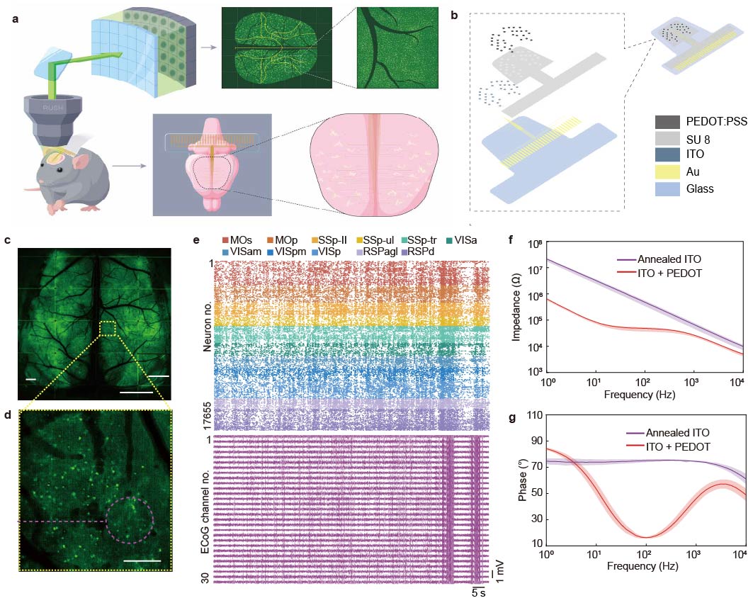
Figure 1: The Cortex-wide Optical-Electrical Recording System CODE
CODE combines a transparent ECoG array with a large-field-of-view single-cell calcium imaging platform, enabling simultaneous acquisition of large-scale neuronal activity and multi-regional electrophysiological signals across the mouse dorsal cortex. This system provides an observational basis for dissecting the cross-scale neural organization of burst-suppression.
Key Finding 1
Under deep anesthesia, the brain does not switch on/off as a whole cortex
━━━━
Using the CODE system, the team simultaneously recorded cortex-wide ECoG signals and calcium activity from tens of thousands of neurons in isoflurane-anesthetized mice. As anesthesia deepened, a typical burst-suppression rhythm emerged in the ECoG signals, with high-amplitude bursts alternating with low-activity suppression phases. At the same time, neuronal calcium activity showed clear state-dependent changes.
Further analysis of neuronal activity patterns revealed two distinct neuronal populations: one preferentially active during bursts, and the other preferentially active during suppression phases. This finding indicates that “suppression” in ECoG signals does not mean complete neuronal silence across the cortex. Even during macroscopically flat phases, more dispersed and asynchronous neuronal activity persists. In other words, bursts and suppression are not simply different activity levels of the same neuronal population; instead, they reflect the alternating organization of distinct neuronal ensembles across brain states.
This finding revises the intuitive view of burst-suppression. Under deep anesthesia, the brain does not simply turn “on” or “off” as a whole. Rather, cortical activity is organized by finer divisions of neuronal populations and their dynamic succession across states.
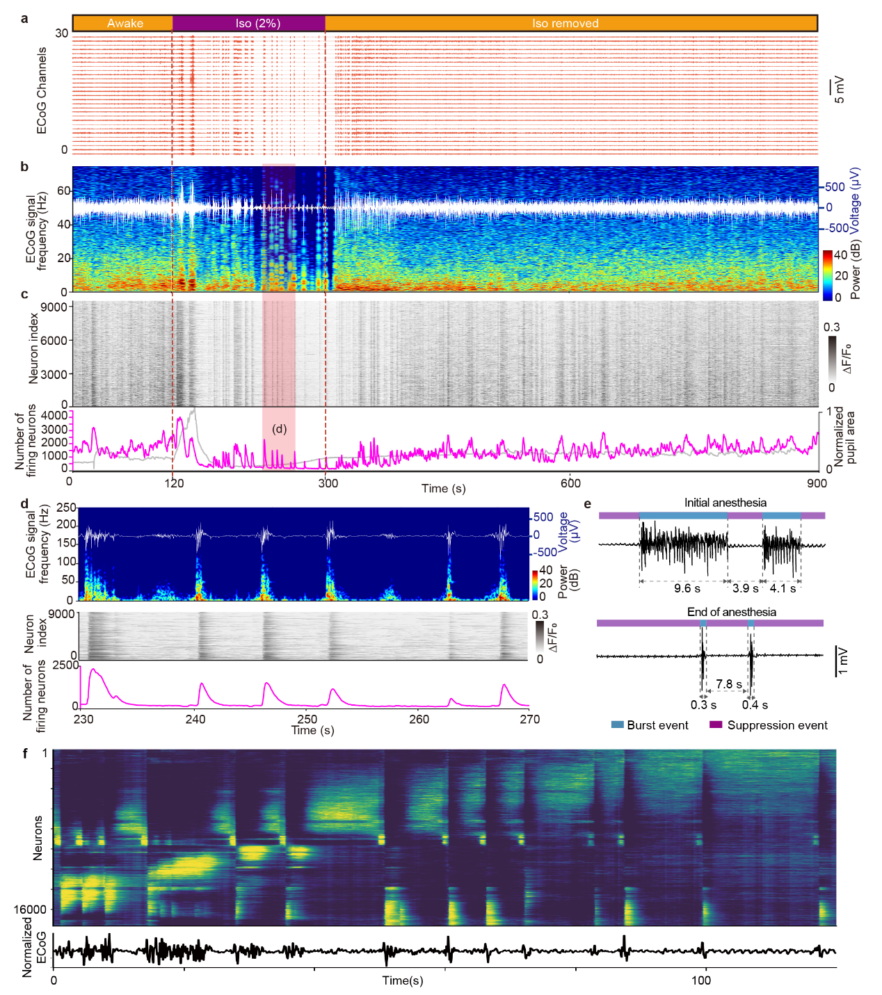
Figure 2: Alternating Activation of Two Neuronal Populations During Burst-Suppression
The study identifies two neuronal populations: burst-associated neurons and suppression-associated neurons. These populations are preferentially active during burst and suppression phases, respectively, and alternate with the burst-suppression rhythm. This indicates that the brain state is not governed by a simple global synchronous switch, but instead has a clear neuronal population-level organization.
Key Finding 2
Bursts and suppression are not repeated replays of a fixed template
━━━━
The team further tracked how these two neuronal populations changed throughout the course of anesthesia. The results showed that burst-associated and suppression-associated neurons were not fixed, unchanging groups. As anesthesia progressed, burst duration gradually shortened, and the number of active neurons participating in bursts decreased. Suppression phases became longer, while the number of neurons involved in suppression-related activity also declined. Cross-event tracking further revealed that each burst or suppression event involved both newly recruited neurons and neurons that had participated in previous events. Neurons recruited more recently were more likely to reappear in subsequent events.
These results suggest that burst-suppression is not the mechanical repetition of a fixed neuronal template, but a continuously evolving process of neuronal population reorganization.
In terms of temporal organization, burst-associated neurons typically reached peak activity early in the burst and showed stronger synchrony. In contrast, suppression-associated neurons reached their activity peaks later during the suppression phase, with activity distributed more broadly over time. Functional connectivity analysis further showed stronger correlations and network connectivity among neurons during bursts, whereas neuronal activity during suppression was weaker and more dispersed. These differences help explain why burst phases appear as high-amplitude waveforms in macroscopic ECoG, while suppression phases appear as low-amplitude or nearly flat signals.
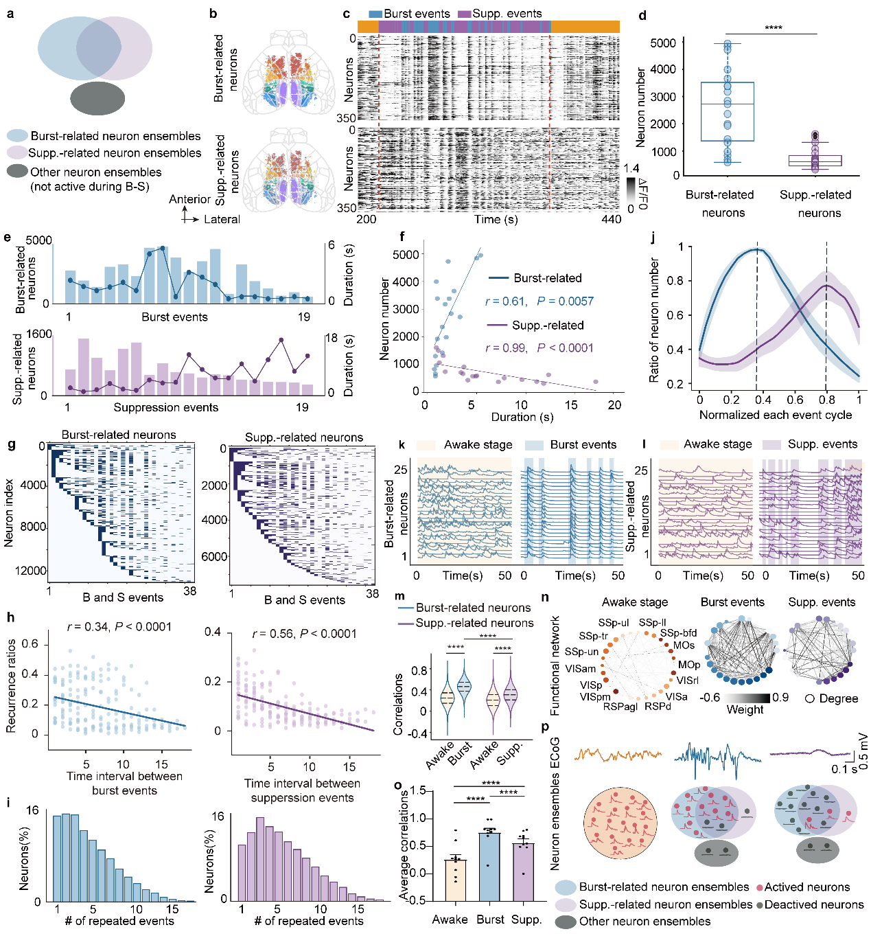
Figure 3: Dynamic Recruitment and Functional Connectivity of Neuronal Populations During Burst-Suppression
Burst-associated neurons are rapidly and synchronously activated early in the burst, forming stronger functional connections. Suppression-associated neurons are recruited later and in a more dispersed manner, with weaker connectivity. The distinct organizational patterns of these two neuronal populations together shape the waveform differences between burst and suppression in macroscopic ECoG.
Key Finding 3
State transitions have spatiotemporal structure rather than occurring as an instantaneous global switch
━━━━
If burst-suppression is not a simple global on-off switch, then the transition from suppression to burst may also have an observable spatiotemporal structure.
The team further analyzed cortical activity during the suppression-to-burst transition. Using spatial grouping and temporal alignment of multi-channel ECoG signals, they found that electrical activity across cortical regions did not change simultaneously around burst onset. Instead, it followed a stable propagation trajectory: burst activity typically first appeared in bilateral lateral sensory cortices and then propagated toward medial and anterior motor-related areas.
Single-cell calcium activity closely matched this electrophysiological propagation pattern. Neurons near the lateral sensory cortices showed broader activity peaks with more dispersed timing, whereas neurons in medial motor-related areas showed sharper activity peaks that were more tightly aligned with burst onset. Cross-modal correlation analysis between optical and electrical signals further revealed that synchrony between ECoG and calcium activity gradually increased during propagation, indicating progressive synchronization during the propagation process.
This finding further challenges the simplified view of burst-suppression as a globally synchronous event. Bursts do not cover the entire cortex simultaneously from the beginning. Instead, as activity propagates from sensory-related areas toward motor-related areas, stronger neuronal synchrony and cross-modal coupling gradually emerge. Thus, even under deep anesthesia, cortical activity is not entirely random or homogeneous, but retains resolvable spatiotemporal organization.
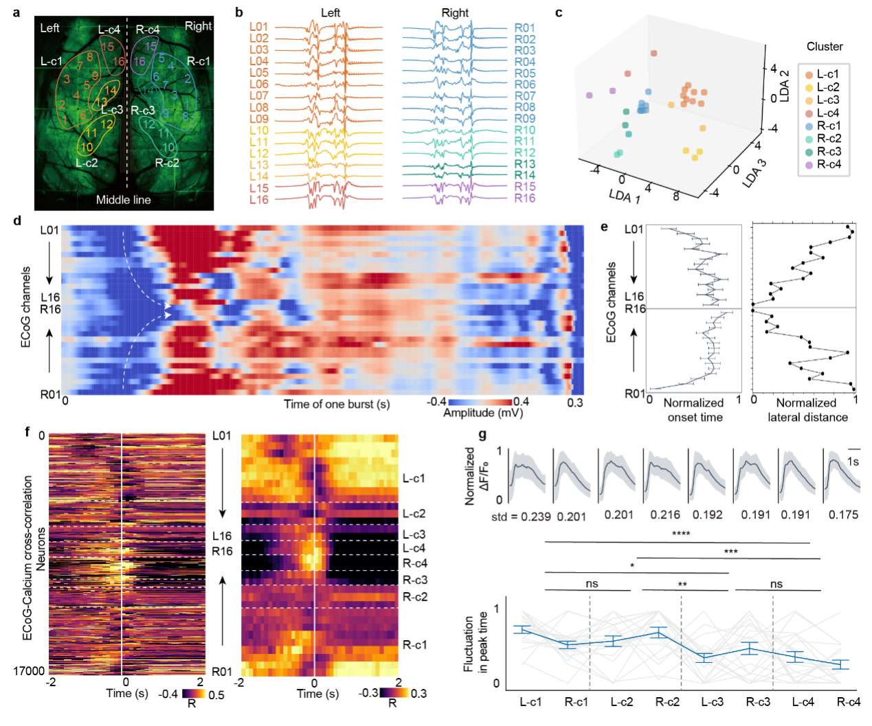
Figure 4: Ordered Propagation of Cortical Activity During the Suppression-to-Burst Transition
Multi-channel ECoG and single-cell calcium imaging together show that, during the suppression-to-burst transition, cortical activity does not emerge synchronously across the entire cortex. Instead, it exhibits regional timing differences and dynamic organization, suggesting that cortical state transitions under deep anesthesia retain resolvable spatiotemporal structure.
Cross-modal Analysis
When can macroscopic electrical signals “read out” single-cell activity?
━━━━
After revealing the neuronal population organization and spatiotemporal dynamics of burst-suppression, the team further asked: To what extent can macroscopic ECoG signals reflect underlying single-neuron activity?
To address this question, the team established a cross-modal prediction framework. First, temporal and spectral features were extracted from multi-channel ECoG signals. At the same time, shared variance component analysis (SVCA) was used to obtain a low-dimensional representation of calcium activity from tens of thousands of neurons. Reduced-rank regression was then applied to build a prediction model from ECoG features to calcium-derived shared variance components, which were further used to reconstruct population neuronal activity.
The results showed that the ability of ECoG to predict calcium activity was strongly brain-state dependent: it was lowest in the awake state, improved during suppression, and reached its highest level during bursts.
This finding indicates that macroscopic electrical signals do not reflect fine-scale neuronal activity in the same way across all brain states. When neuronal activity is more synchronous, network coupling is stronger, and population dynamics are lower-dimensional, ECoG can more readily capture the corresponding population activity structure. In contrast, during awake states, neural activity is more complex and higher-dimensional, making the correspondence between macroscopic electrical signals and single-cell activity relatively weaker.
These findings provide a quantitative perspective for understanding the interpretability of macroscopic electrical signals. ECoG or EEG should not be regarded simply as a direct “average” of neuronal activity. Instead, the fine-scale neural structures they reflect — and the extent to which they reflect them — may vary substantially across brain states.
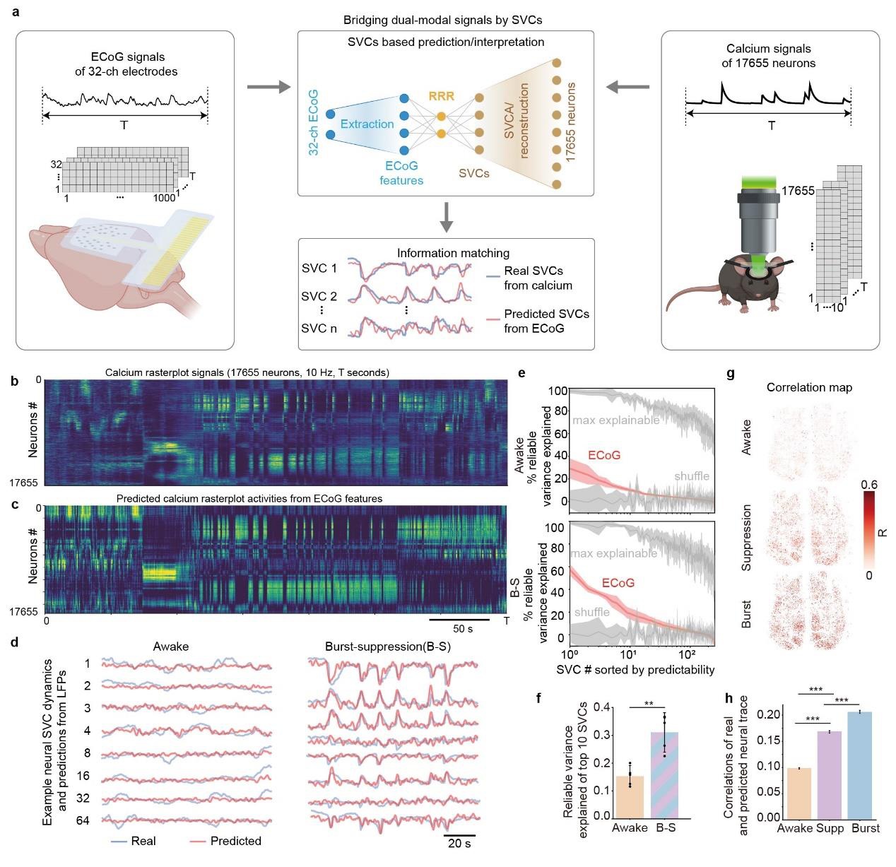
Figure 5: Cross-Modal Correlation and Prediction Based on Shared Variance Components
Using shared variance components and a cross-modal prediction model, the study links macroscopic ECoG features to calcium activity from tens of thousands of neurons. The results show that ECoG prediction of calcium activity is strongly brain-state dependent, with the highest predictive power during bursts, intermediate predictive power during suppression, and the lowest predictive power in the awake state.
Summary and Outlook
Burst-suppression is an organized cortical dynamic state
━━━━
Through the CODE system, this study placed calcium activity from tens of thousands of single neurons and multi-regional ECoG recordings on the same timeline, systematically dissecting the cross-scale organization of burst-suppression under deep anesthesia.
The study revealed four key findings.
First, bursts and suppression are not simple intensity variations of the same neuronal population. Instead, they involve distinct neuronal ensembles that show preferential activity during different phases.
Second, burst phases exhibit stronger synchrony and higher functional connectivity, whereas suppression phases show more dispersed and asynchronous activity.
Third, the suppression-to-burst transition has resolvable cortical spatiotemporal structure, rather than occurring as a simple global synchronous switch.
Fourth, the ability of macroscopic ECoG signals to explain underlying single-cell population activity depends on brain state. This interpretability is strongest during highly synchronous bursts and weaker during the complex, high-dimensional awake state.
Together, these findings indicate that burst-suppression is not a homogeneous, passive silencing of the entire cortex. Rather, it is a complex cortical dynamic state shaped by neuronal population recruitment, changes in functional connectivity, and cross-regional spatiotemporal organization.
Outlook
The integrated optical-electrical recording capability demonstrated by CODE provides new tools and perspectives for future research
━━━━
• Dissecting cross-scale principles of brain state transitions: By simultaneously capturing high-speed electrophysiological rhythms and microscale neuronal population activity, CODE may help further investigate dynamic transitions across brain states such as anesthesia, sleep, coma, and epilepsy.
• Developing interpretable brain-state monitoring methods: By establishing cross-modal correspondences between ECoG and single-cell population activity, this study provides a quantitative framework for understanding the neural basis of macroscopic electrical signals across different states of consciousness.
• Extending to cell-type, neuromodulatory, and disease-model studies: Future integration with higher-density transparent electrode arrays, wider-bandwidth electrophysiological recording, and cell-type-specific imaging may further improve the resolution of cortical dynamics analysis and extend the platform to complex cognitive tasks and neurological disease models.
Guihua Xiao, Associate Research Fellow at the National Research Center for Information Science and Technology, Tsinghua University; Mo Yang, Ph.D. candidate in the Department of Biomedical Engineering; and Lingbo Li, Postdoctoral Fellow in the Department of Automation, are co-first authors of this article. Associate Professor Xiaochuan Dai (Department of Biomedical Engineering), Academician Qionghai Dai (Department of Automation), Associate Professor Jiamin Wu (Department of Automation), and Associate Research Fellow Guihua Xiao (National Research Center for Information Science and Technology) are co-corresponding authors. Collaborators also include the research group of Bo Hong (Department of Biomedical Engineering, Tsinghua University) and the research group of Ting Lei (Department of Materials Science and Engineering, Peking University).
Original article link: https://www.nature.com/articles/s41467-026-72454-0
Reference: [1] Fan, J. T. et al. Video-rate imaging of biological dynamics at centimetre scale and micrometre resolution. Nature Photonics 13, 809 (2019). https://doi.org:10.1038/s41566-019-0474-7.