1. Background
Brain-computer interfaces, implantable neural electrodes, and flexible wearable devices have attracted growing attention in recent years. These cutting-edge technologies are moving from the laboratory toward everyday use, but long-term implantation brings a new set of challenges. The environment inside the body is highly complex. Biofluids continuously wash over the device, ions repeatedly enter and leave the coating, and the electrode must endure sustained electrical pulses. Whether these devices can operate reliably over time often depends less on the impressive conductivity reported at the outset than on a simple but crucial question: can the functional coating stay firmly attached to the electrode surface? Among the many candidate materials, poly(3,4-ethylenedioxythiophene) (PEDOT)-based polymers have drawn particular interest because they can conduct both electrons and ions. Yet their long-term use has revealed a clear weakness. During chronic operation in the body, PEDOT-based coatings may crack at the interface, delaminate, or even detach completely. Such failures directly affect device safety and the prospects for clinical translation.
To address this problem, a team led by Xiaochuan Dai at Tsinghua University's School of Biomedical Engineering published a comprehensive review in ACS Nano titled “Seaming the Bioelectronic Interface: Mechanisms, Strategies, and Validation Standards for Durable Poly(3,4-ethylenedioxythiophene)-Based Coating Adhesion.” The article systematically reviews the origins of adhesion failure in PEDOT-based coatings, strategies for strengthening the interface, and approaches to standardized validation. It offers a structured framework for addressing long-term stability in brain-computer interfaces, implantable electrodes, flexible wearables, and other bioelectronic systems.
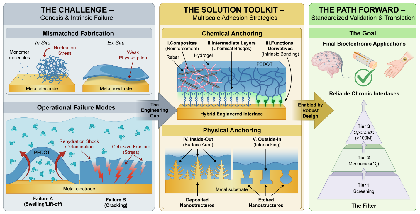
Figure 1. A unified framework for PEDOT-based coating interface engineering, from failure mechanisms and reinforcement strategies to standardized validation. The left panel shows the typical failure modes associated with in situ growth and ex situ deposition. The center summarizes chemical and physical anchoring strategies. The right presents a three-tier validation framework that provides a systematic path from laboratory testing to the clinical translation of durable bioelectronic interfaces.
2. Two Failure Mechanisms: Stress Accumulation and Rehydration Shock
Before the problem can be addressed, its underlying mechanisms must first be understood. This review shows that delamination of PEDOT-based coatings is not incidental, but arises from two structural factors introduced during fabrication. Broadly speaking, PEDOT-based coatings are formed through one of two approaches: in situ growth or ex situ deposition.
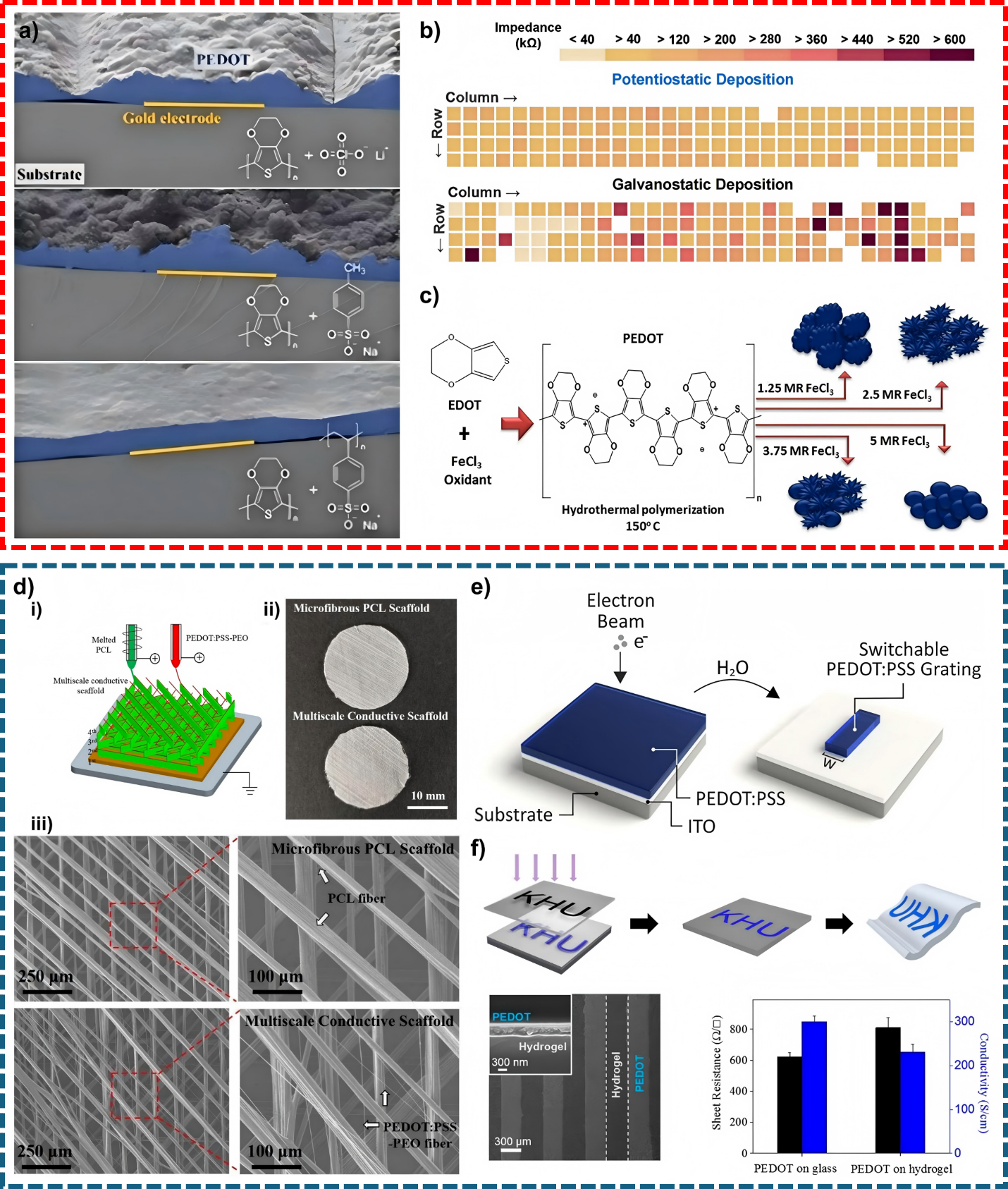
Figure 2. Two representative routes for forming PEDOT-based coating interfaces: in situ growth and ex situ deposition. Representative in situ growth methods are shown in the red box at the top, while representative ex situ deposition methods are shown in the blue box below.
In situ growth refers to polymerizing EDOT monomers directly on the electrode surface through methods such as electropolymerization, chemical oxidative polymerization, and vapor-phase polymerization. Because the polymer forms on the substrate, the coating can closely follow the underlying surface topography. However, the reaction is driven mainly by oxidation of the monomer rather than by a chemical reaction between the growing polymer and the substrate. The interface is therefore often held together mainly by weak physical adsorption. The situation resembles building a house before laying a solid foundation. During long-term operation, ions and water molecules repeatedly move into and out of the coating. The material undergoes pronounced volumetric “breathing,” alternating between swelling and contraction. As these cycles continue, mechanical stress accumulates at the interface. Once it exceeds the strength of the physical adhesion, cracks can form and the entire coating may eventually peel away. The review describes this failure mode as “stress accumulation.”
Ex situ deposition begins with a preformed PEDOT solution, ink, or composite, which is then transferred to the target substrate through spin coating, spray coating, printing, lithographic patterning, or related processes. This route is flexible, suitable for large-area fabrication, and widely used in flexible and wearable electronics. Because polymer synthesis and film deposition occur as separate steps, however, the interface often depends on weak interactions such as van der Waals forces. The process is much like a dried sponge. The coating may look compact and firmly attached in the dry state, but once it is re-exposed to biofluids or a biofluid-like environment, hydrophilic components such as PSS rapidly absorb water and swell. This creates a sudden osmotic stress near the interface. The film can begin to lift from the edges and eventually detach as a whole. This failure mode is known as “rehydration shock.”
Two Failure Mechanisms, Two Design Responses
The key difference between in situ growth and ex situ deposition lies in their dominant modes of failure. In situ-grown coatings are more vulnerable to mechanical stress that builds up over long periods, whereas ex situ-deposited coatings are more likely to fail rapidly when they encounter water again. This distinction offers a clearer way to evaluate the reliability of PEDOT-based coatings. Durability cannot be judged by adhesion strength alone. The fabrication route and the failure mode most likely to occur under realistic conditions must also be considered so that the reinforcement strategy can be matched to the actual risk.
3. Two Complementary Strategies: Chemical and Physical Anchoring
With these failure mechanisms identified, the review categorizes adhesion-enhancement strategies for PEDOT-based coatings into two broad approaches: chemical anchoring and physical anchoring. These approaches address the distinct mechanisms associated with the two fabrication routes described above.
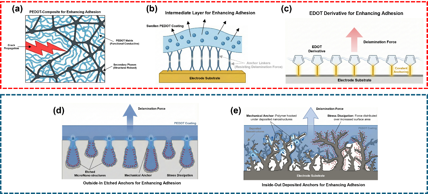
Figure 3. Schematic illustration of chemical and physical anchoring strategies. The red box at the top presents three representative mechanisms for chemical anchoring, while the blue box below shows two complementary routes for physical anchoring.
Chemical anchoring aims to replace or reinforce weak physical adsorption with stronger and more specific interactions. The review groups these approaches into three categories. The first uses composite reinforcement. Carbon nanotubes or adhesive hydrogel networks are incorporated into PEDOT and act much like rebar in concrete, redistributing stress and slowing crack propagation. The second introduces an intermediate linker layer, such as a silane, polydopamine (PDA), or a conductive polymer. This layer acts like double-sided tape in a molecular sandwich. One side binds to the substrate, while the other entangles or reacts with PEDOT. The third uses functionalized EDOT derivatives. By introducing reactive groups into the EDOT monomer, as in EDOT-NH2, the polymer-forming layer can establish covalent attachment to the substrate during growth. This advanced form of “in situ grafting” addresses the weak boundary created when the polymer is formed first and attached only afterward.
Physical anchoring does not depend on forming new chemical bonds. Instead, it reshapes the substrate surface so that PEDOT becomes mechanically locked in place, much like a mortise-and-tenon joint. The review highlights two complementary routes. In “inside-out” deposition, high-surface-area dendritic structures, such as “fuzzy gold” or nanostructured iridium oxide, are first created on the electrode. PEDOT then grows around and through these structures like a vine, increasing the contact area and distributing stress. In “outside-in” etching, laser or chemical treatments create re-entrant micropores or grooves in the substrate. After PEDOT fills and solidifies within these features, it becomes mechanically interlocked and is difficult to remove as a continuous film, even under substantial stress.
These two paradigms are not mutually exclusive. Bioelectronic interfaces designed for long-term implantation often need both molecular-scale bonding strength and macroscopic mechanical interlocking.
4. Establishing a Standard: The Proposed “Three-Tier Validation Pyramid”
PEDOT adhesion research has long lacked a unified evaluation framework. Some studies use tape tests, whereas others rely on ultrasonication; some report stability following cyclic voltammetry, whereas others monitor changes in impedance. Differences in test conditions, failure criteria, and reporting metrics make it difficult to compare strategies directly or to assess their performance within a systematic, staged validation framework for medical devices. To address this gap, the authors propose a “3-Tier Validation Hierarchy” that links material-level adhesion assessment with a progressive device-validation framework:
Tier 1 (Rapid Qualitative Screening: The Gatekeeper)
Tape tests, ultrasonication, and short-term soaking provide rapid initial screening. They quickly rule out interfaces with obvious weaknesses and reduce the cost of more demanding follow-up tests.
Tier 2 (Quantitative Mechanics: Core Metrics)
Interfacial fracture energy, shear strength, crack density, local modulus, and related measurements turn the vague impression that a coating “seems firmly attached” into quantitative data that can be compared across studies.
Tier 3 (Operando Durability: The Preclinical Gold Standard)
Long-term electrical pulse cycling, including tests exceeding 100 million pulses, is combined with accelerated aging, electrochemical monitoring, histological evaluation, and functional recording. Together, these tests allow candidate materials to undergo as comprehensive a rehearsal as possible of the challenges they may later face in the body.
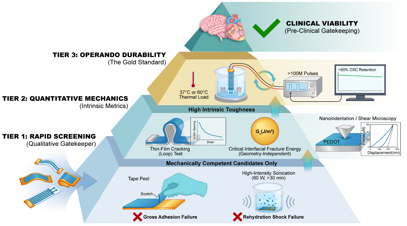
Figure 4. The three-tier validation hierarchy for adhesion of PEDOT-based coatings.
The value of this framework lies not only in moving adhesion evaluation beyond qualitative judgments of whether a material remains attached, but also in enabling staged assessment and more meaningful comparison across studies. The authors further suggest that the framework may serve as a systematic reference for the development of relevant industry and association standards in China, while facilitating alignment with internationally recognized validation practices. By supporting more consistent and standardized evaluation of bioelectronic interface reliability, it could help inform future product registration and clinical translation.
5. Summary and Outlook: From Interfacial Separation to Interfacial Integration
This review does more than summarize the current state of the field; it also considers future directions for PEDOT-based interfaces. The authors suggest that next-generation bioelectronic interfaces may need to progress from “static attachment” toward dynamically adaptive systems. They outline six directions for further development.
First, chemical and physical anchoring may be combined to integrate macroscopic mechanical interlocking with microscopic covalent bonding. Second, failure assessment may shift from post-failure analysis toward real-time monitoring. In situ techniques, including scanning electrochemical cell microscopy (SECCM), Brillouin microscopy, surface-enhanced Raman spectroscopy (SERS), and surface-enhanced infrared absorption spectroscopy (SEIRA), could help identify early indications of interfacial softening before crack propagation. Third, AI-assisted materials design could combine density functional theory (DFT) calculations with graph neural networks to predict the relative durability of interface designs before physical prototypes are fabricated. Fourth, self-healing interfaces based on dynamic covalent chemistry may enable the repair of microcracks. Fifth, reversible connections could facilitate the removal of implanted devices with minimal tissue disruption. Finally, translation toward scalable manufacturing will require multistep fabrication procedures to be adapted into stable processes compatible with continuous roll-to-roll production.
Toward More Integrated Bioelectronic Interfaces
The review notes that addressing the physicochemical mismatch between organic polymers and inorganic substrates is important for the long-term development and clinical translation of bioelectronic devices. Its framework for analyzing coating failure may also be relevant to other electrochemical technologies that involve interfacial degradation, such as anode pulverization in energy-storage devices. More broadly, the authors emphasize that improving bioelectronic interfaces involves more than increasing adhesion strength. It also requires reducing mechanical and chemical discontinuities between living tissue and engineered materials, thereby supporting more stable integration at the interface.
This perspective may also have implications beyond individual laboratory studies. The authors suggest that the proposed 3-Tier Validation Hierarchy could serve as a scientifically grounded reference for the development of future standards for bioelectronic devices in China, while facilitating alignment with internationally recognized validation practices. By supporting more consistent evaluation of interface reliability, the framework may help inform future device development, product registration, and clinical translation.
Author Information
The review was published in ACS Nano. Its co-first authors are Kai San Chan, a 2024 graduate of School of Biomedical Engineering and currently a PhD student in the Department of Biomedical Engineering at the National University of Singapore; and Yifan Guo, a 2025 graduate of Tanwei College and currently a PhD student at the University of Chicago’s Pritzker School of Molecular Engineering. Xiaochuan Dai, Associate Professor at Tsinghua University’s School of Biomedical Engineering, is the corresponding author.
Original article: https://doi.org/10.1021/acsnano.6c05215