Recently, Professor Kexin Yuan’s team from the School of Biomedical Engineering, Tsinghua University published a research article in Cell, titled “VIVIT: Resolving trans-scale volumetric biological architectures via ionic glassy tissue.” This study introduces and validates a novel tissue processing method, named VIVIT, which enables high-fidelity three-dimensional imaging of biological tissues in a glassy state. VIVIT overcomes three long-standing bottlenecks in tissue clearing technologies, achieving transparency without deformation, preventing fluorescence attenuation, and enabling damage-free cryopreservation and sectioning, thus opening new avenues for basic biological research, pathology, and AI-assisted diagnosis, including applications in neuroscience.

Three-dimensional tissue architectures contain rich biological information essential for understanding physiological functions and disease mechanisms. Traditionally, however, this information has relied heavily on 2D histological section samples are cut into dozens or even hundreds of thin slices that are then stitched together. This process is labor-intensive and prone to structural deformation or rupture, often leading to spatial information loss and misinterpretation.
To address these limitations, tissue clearing techniques have emerged, using chemical methods to render tissues optically transparent and enable deep imaging without physical slicing. However, existing approaches face persistent challenges, such as tissue expansion or shrinkage, loss of fluorescence signals, and fragility during freezing, making it difficult to achieve an optimal balance between structural stability, signal preservation, and staining compatibility.

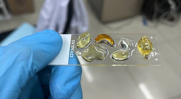
VIVIT provides an innovative solution to these challenges. Unlike conventional clearing methods that focus solely on transparency, VIVIT emphasizes both optical clarity and preservation of native structural and fluorescent integrity. Through the use of a custom-developed high–refractive index ionic liquid, the team successfully transformed opaque biological tissues into a “glassy state” at low temperature—a stable and transparent condition with less than 1% structural deformation. Even complex and delicate samples such as brain tissues maintained their original architecture after VIVIT processing, allowing for clear visualization of subcellular structures, including synaptic connections. The study demonstrated this across rodent, non-human primate, and human brain tissues, highlighting the broad applicability and robustness of the technique.
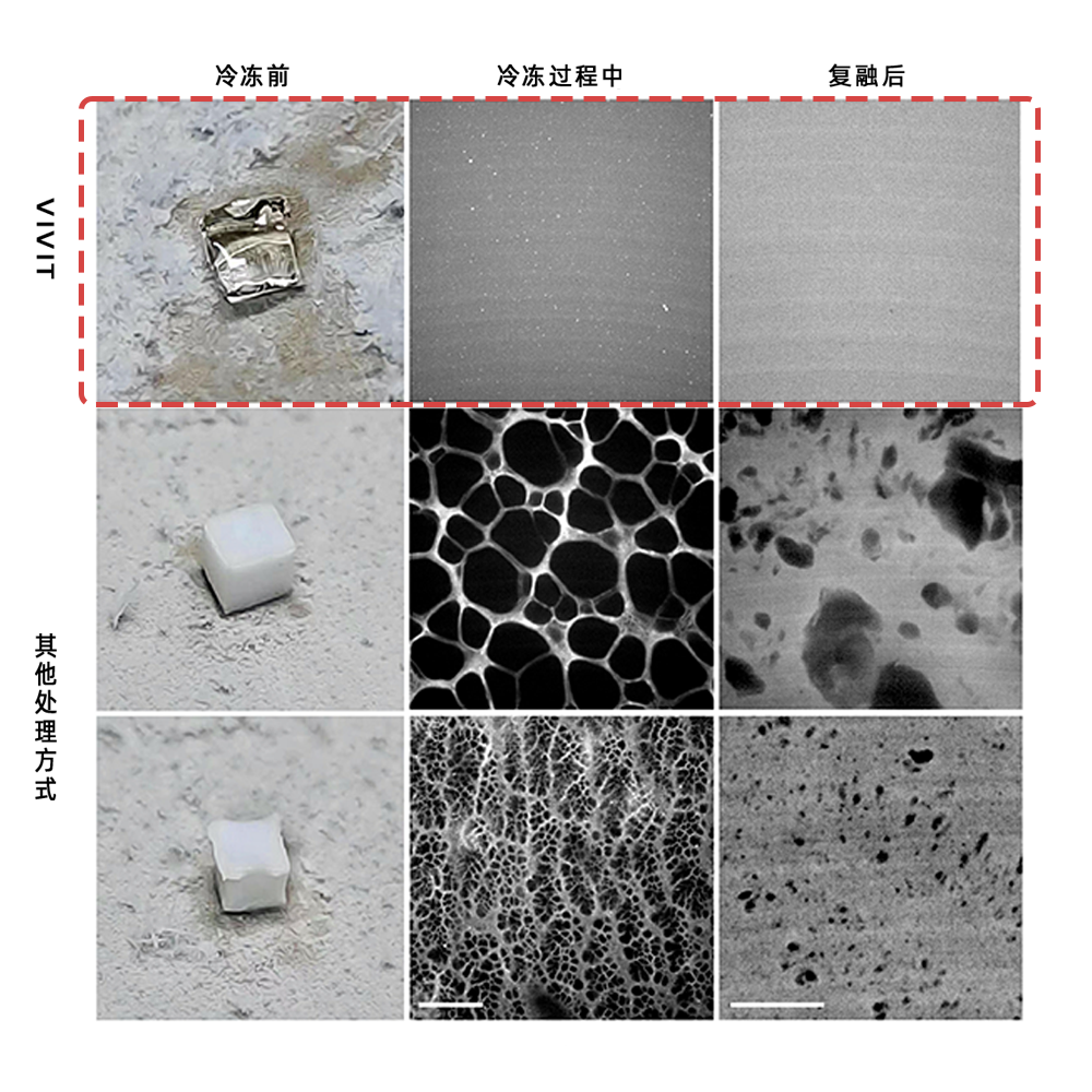
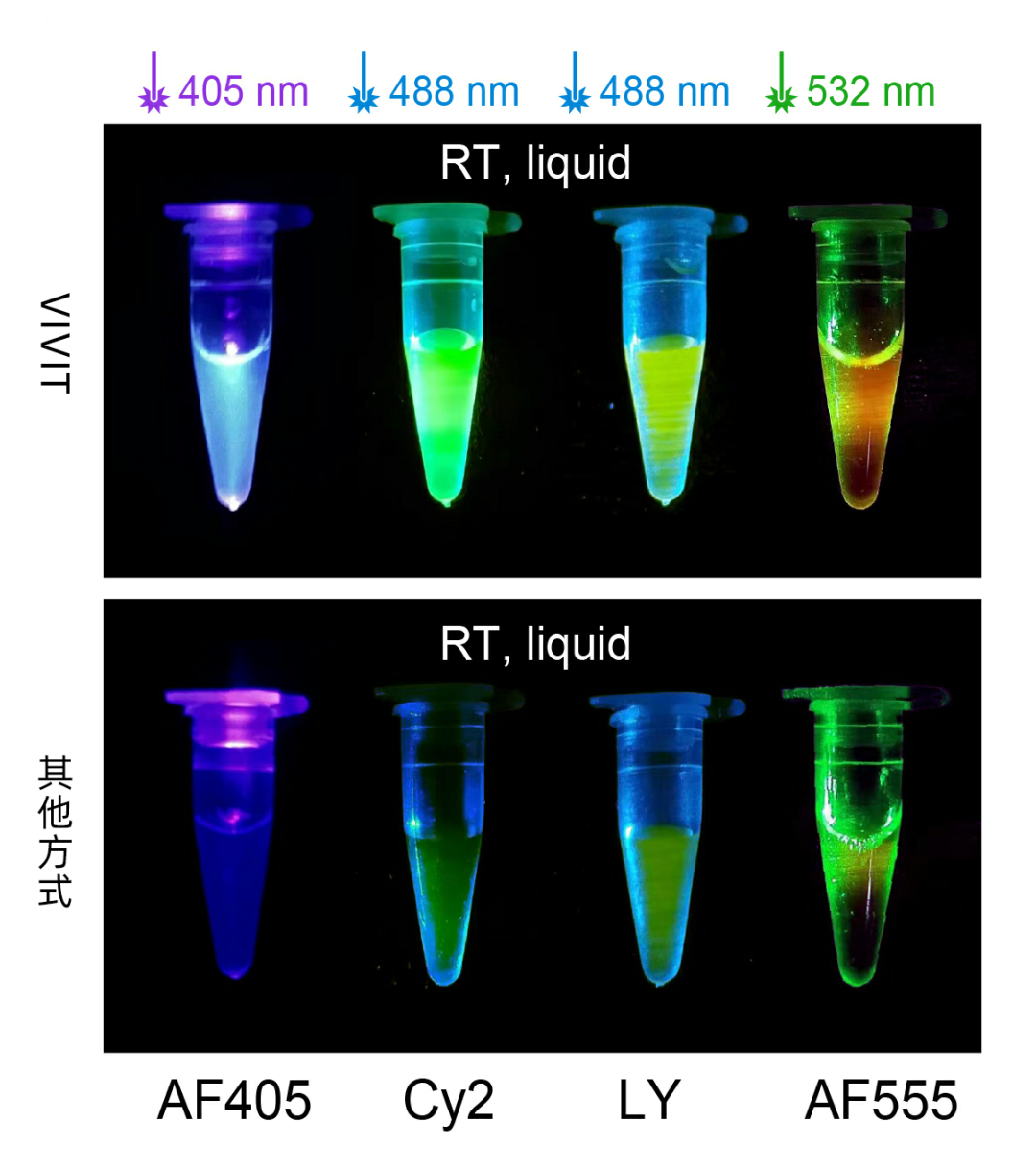
VIVIT’s core innovations extend beyond achieving optical transparency, it also ensures signal preservation and functional usability. After ionic-liquid treatment, the fluorescence intensity of multiple commonly used dyes was enhanced by 2–30 times, enabling the detection of previously undetectable weak signals. Furthermore, owing to its glassy-state physical properties, VIVIT overcomes the limitations of traditional cryopreservation. Samples can be stored at −80 °C long-term without ice-crystal formation or mechanical damage, thus enabling structurally intact cryosectioning and super-resolution imaging. This combination of high structural fidelity and signal stability establishes a solid foundation for acquiring and reconstructing trans-scale 3D biological data.
Leveraging these advantages, Professor Yuan’s team quantitatively mapped the relationship between the sensory modality preferences of thalamic neurons (microscale) and their projection targets across the entire brain (meso- and macroscale) in mice—marking the first accurate input–output linkage at the single-neuron level globally. This breakthrough represents a major step forward in neural circuit research, offering new opportunities to uncover the mechanisms underlying brain function.
Importantly, VIVIT-treated tissues remain suitable for multiple rounds of immunostaining, with each round maintaining clear fluorescence and stable structural integrity. This allows researchers to sequentially identify multiple molecular targets within the same sample, greatly enriching spatial information. Combined with the team’s custom reconstruction algorithm, TARS, VIVIT supports image registration and stitching of serial tissue sections, enabling the creation of 3D maps from subcellular to whole-organ levels and facilitating cross-scale biological reconstruction.
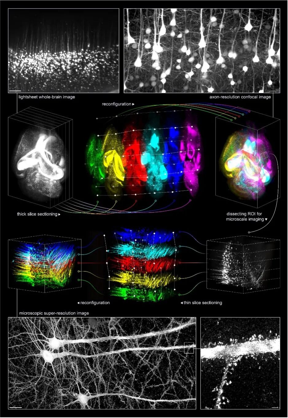
From tissue clearing to multiplexed labeling and 3D reconstruction, VIVIT establishes a complete technological pipeline covering every step from sample processing to spatial structural analysis. It provides a systematic solution for high-resolution, trans-scale spatial data acquisition and tissue modeling.
In the future, Professor Yuan’s team will continue to advance the application of VIVIT in neuroscience, while expanding its use across broader fields of life sciences, precision medicine, and intelligent diagnostics, unlocking the full scientific and clinical potential of spatial structural information.
Article Link: https://doi.org/10.1016/j.cell.2025.07.023
About the Research Team
Professor Kexin Yuan’s group at the School of Biomedical Engineering, Tsinghua University, conducts interdisciplinary, cross-scale, and cross-species research focused on two closely related but distinct themes: The integration mechanisms of multimodal sensory information, and the pathological mechanisms underlying sensory disorders. In recent years, the team has published a series of original works as corresponding author in leading international journals, including Cell (2025), Neuron (2023), IEEE Transactions on Pattern Analysis and Machine Intelligence (2024), Journal of Neuroscience (2024), and Cerebral Cortex (2019).